
A mole is a common skin growth, characterized by a circular shape and a darker pigment than the surrounding skin. Also referred to as “melanocytic nevus”, moles are created by groups or clusters of melanocytes in the skin.
Melanocytes are cells that produce melanin, the skin’s natural pigment. These cells exist evenly throughout the body, but they sometimes group together, which creates hyperpigmentation and the formation of common moles.
Atypical moles, also called dysplastic nevi, are moles with ‘irregular’ characteristics, specifically in size, shape, border, color, or texture. Although atypical moles are benign (non-cancerous), their presence is linked to an increased risk of melanoma (the most serious form of skin cancer).
People with 10 or more atypical moles have 12x the risk of developing melanoma. Atypical moles resemble melanoma, which is why mole removal is so critical.
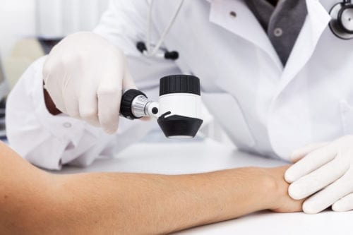
If your dermatologist identifies an atypical mole during your annual skin cancer check, he or she will suggest a mole removal procedure to perform a biopsy of the mole. During a mole biopsy, the doctor examines the cells in the mole to determine whether the mole is malignant or benign.
How to Identify an Atypical Mole
It’s normal for moles to vary slightly in appearance, but there are a few characteristics and features to look out for. Dermatologists use these ABCDE warning signs to help diagnose skin cancer, by identifying atypical potentially-cancerous moles.
An (Asymmetry): Most common, benign moles have a symmetrical shape. These moles can be (figuratively) cut down the middle, and the two halves will resemble each other identically. Atypical moles and cancerous moles often have an asymmetrical shape with two, non-identical halves.
B (Border): Similar to the asymmetrical shape, the border of an atypical mole is often jagged or uneven, with edges that ‘bleed’ into the surrounding skin pigment. Common moles, on the other hand, have smooth, even borders with distinct edges.
C (Color): The color of an atypical mole may be uneven with areas of light brown pigment and areas of darker black/brown pigment. If an atypical mole starts to grow and develops areas of red or blue pigment, this is indicative of melanoma.
D (Diameter): If an atypical mole grows to the size of a pencil eraser (approximately 6 mm, or ¼ inch in diameter), this is a sign that the mole is cancerous. It’s important to have moles removed and biopsied before they grow larger and lead to other potential complications.
E (Evolving): Any changes in the appearance of a mole should be noted and brought to a dermatologist immediately. Evolution in the mole’s size, shape, color, border, elevation, or texture can indicate melanoma. If any new symptoms (like bleeding, crusting, or itching) develop, this can also indicate skin cancer.
Although atypical moles are not cancerous, atypical moles have a similar appearance to cancerous moles. Performing self-examinations regularly, using the ABCDE warning signs as a guideline, is critical for detecting skin cancer before it develops further.
Every month, examine your skin in the mirror and pay close attention to your moles. Assess the border, color, size, and shape of every mole and stay conscious of any changes or evolution over time.
Immediately notify your dermatologist of any changes in your moles, because skin cancer is highly-treatable if it’s caught in the early stages.
Risk Factors for Melanoma
There are a few risk factors that increase a person’s chances of developing melanoma.
- Fair skin
- Light eyes
- Light hair
- Freckles
- Photosensitivity (often due to a medical condition, medications or genetics)
- Many moles on the body
- Family history of skin cancer
- Skin that burns easily in the sun
Atypical Mole Removal Procedures
If your dermatologist identifies a suspicious atypical mole on your body, there are a few treatment options available to remove the mole, examine the skin cells and treat skin cancer (if necessary). The first step to treat melanoma is to remove the mole (or an area of the mole) and examine the skin cells.
Any of the following biopsies may be performed to examine the skin cells:
Shave biopsy
During a shave biopsy procedure, a thin layer of skin in the affected area is removed with a small blade (similar to a razor). A local anesthetic is typically applied to numb the area before removing the skin.
Stitches are not necessary to repair the skin after a shave biopsy. A scab will form over the wound, which generally heals the skin within a week or so.
Punch biopsy
Punch biopsies remove a larger area of skin than shave biopsies. During these biopsies, a rounded blade is rotated around the area to remove a thick skin sample that includes multiple layers of the skin, including the epidermis, dermis, and subcutis.
Because a punch biopsy extracts multiple layers of skin, stitches are sometimes required to close the wound. A bandage will be placed over the area after the mole removal procedure to prevent further bleeding.
Surgical excision biopsy
A surgical excision biopsy procedure involves the removal of an entire piece of skin tissue on and around the mole, by use of a scalpel. Surgical excision biopsies remove the entire skin lesion, down to the subcutis skin layer.
Depending on the depth of skin removed during the mole extraction, stitches may be necessary to close the wound and promote skin healing. A bandage will be placed over the area to stop the bleeding.
Scarring after Mole Removal
The type of scarring that develops after a mole removal procedure will depend on the type of mole removal procedure performed, your age, and the thickness of the skin removed.
Scarring on young people who have high levels of collagen and elastin in their skin generally heals quicker than scars on adults who have naturally declining levels of collagen and elastin production.
Everybody reacts differently to scarring. Depending on factors, such as a person’s immune system reaction, the amount of blood supply in the area of the wound, and the amount of sun exposure to the affected area, scars may be unnoticeable and blend in with the surrounding skin over time, or they may form as elevated keloid or hypertrophic scars.
Compared to excision biopsies and punch biopsies, shave biopsies typically remove the least amount of skin, so these procedures don’t require sutures or stitches to close the wound. The scarring that develops after a shave biopsy often appears as a light pink patch of skin where the mole once was.
An experienced dermatologist will use a minimally-invasive approach to remove the mole and leave as little scarring behind as possible. Scars from excision biopsies and punch biopsies can require more time to heal fully. Some people’s scars will lighten and blend in with the surrounding skin, while other people’s wounds may give rise to thicker keloid or hypertrophic scars.
Wound Care after Mole Removal
Following a mole removal procedure, you’ll wear a bandage wrapped around the wound for about 24 hours. After 24 hours, you’ll remove the bandage and clean the wound with gentle, soapy water and a soft gauze or Q-tip.
It’s important to rinse the area thoroughly to eliminate leftover soap suds. Dry the skin with another gauze or Q-tip. Apply a thin layer of Vaseline or Aquaphor to the area, and cover the area with a band-aid or bandage. Repeat these steps once a day until you see the wound is healed completely.
You can shower as normal after your mole removal procedure, but don’t submerge your bandage or wound in water. Anytime your bandage gets wet, clean the wound thoroughly (as described above) and replace it with a dry bandage.
Over time, diligent care of your wound will support the skin in healing and recovering fully from the procedure.
Next Steps
Once the mole is successfully removed, the skin sample is sent to a lab for examination. Lab results typically take about a week to be produced, and your dermatologist will call you to notify you of their findings.
If the cells in your mole are healthy and noncancerous, there is no further action necessary. You’ll follow your dermatologist’s post-procedural instructions, and your wound will heal over time.
If the cells in your mole are cancerous, your dermatologist will discuss your treatment options with you. Depending on the specific type of skin cancer and how far it’s developed, your dermatologist may suggest one skin cancer treatment over another.
We advise any person who has a suspicious mole to schedule an appointment with us and visit our exceptional Orlando dermatologists. It’s incredibly important that we examine any potentially cancerous moles, because early detection and treatment of skin cancer can be life-saving.
Treating skin cancer in the early stages has proven highly effective with minimal downtime. If skin cancer (specifically melanoma) progresses to the later stages, it can spread beyond the skin, to the lungs, brain, bones, and lymph nodes.
Our experienced dermatologists are here for you! Our dermatologist is a board-certified dermatologist with specialty training in Mohs surgery for the treatment of skin cancer.
At UCF Health, we are here for your dermatology needs! We want your skin to be in its healthiest condition, and that includes general screenings, exams, and tests for the detection and prevention of skin cancer.
If you’re due for your annual skin cancer check, we encourage you to schedule an appointment today. Through our convenient patient portal, you can view test results and communicate with your doctor to ask questions or express any concerns.
We want to help educate you and provide you with the resources you need to maintain your healthiest skin.