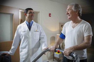
Colloquially known as a stress test, a stress echocardiogram is a non-invasive procedure that measures how your heart, its related blood vessels and arteries are working. The word “stress” as it relates to this cardiovascular test doesn’t in fact mean the typical kind we often think of – irritation related to traffic, work, finances, and relationships. This type of stress relates to physical stress caused by exertion that the heart undergoes during exercise and physical exertion.
The test works by placing an individual on a treadmill or bicycle, then working the heart up to peak cardio levels. The cardiologist will take an ultrasound of the heart while it’s working hard to see how much blood and oxygen it’s receiving while exercising. They will also monitor blood pressure, heart pulses and rhythm, carefully recording the data points for further review.
Stress echocardiography comes into play when an individual has an existing cardiovascular issue or may have cardiovascular concerns related to developing symptoms. The cardiologist may want to rule out cardiovascular issues as they relate to common symptoms that can be indicative of a variety of diseases. Stress tests are ordered for a number of physical signs and symptoms, including but not limited to:
- Chest pain
- Shortness of breath
- Feeling fatigued after minimal physical exertion
- Racing heart beats
- heart palpitations
- History of high blood pressure
How is a Stress Echo performed?
Prior to a stress echocardiogram, a regular (resting) echocardiogram will be performed. The patient lies on his or her left side with the left arm extended. A gel is applied to the chest to help ultrasound waves from the transducer reach the heart. The ultrasound sends information back related to the heart rate and rhythm while the cardiovascular system is in a resting state.
After this, a stress echocardiogram will be performed. The patient can walk on a treadmill or ride a stationary bike. About every three minutes, he or she will be instructed to jog or pedal faster in order to ramp up the heart rate and increase blood pressure. The test continues this way for about 15 minutes, or as long as the test provider feels is necessary to get a clear picture of cardiovascular health.
Dobutamine Stress Echocardiogram
If an individual can’t exercise, he or she will receive an injection of dobutamine, which causes an increase in heart rate and blood pressure for a short period of time. As the heart rhythm is increasing and while it’s at peak rate, the care provider or cardiologist will perform another ultrasound of the cardiovascular system. In this way, he or she can see which parts of the heart are more oxygenated and which are lacking. This can help doctors identify blockages and clogs in arteries and veins surrounding the heart.
Risk Factors
A stress echo is a very non-invasive, safe test and has few complications. Your cardiologist should only recommend a stress test if you are able to withstand the exertion of mild to moderate exercise. Side effects of a stress echo are rare but can include:
- Dizziness
- Chest pain
- Fainting
- An irregular heart beat
- Increase in systolic blood pressure
Types of Stress Echocardiograms
- M-mode: This simplistic type of echocardiogram produces an image that looks more like a tracing than a realistic, 3-D image of the heart’s chambers. An m-mode stress test measures the heart structures and thickness of its walls.
- Doppler: This type of stress test is employed to assess the blood flow as it passes through the chambers and valves. A Doppler test can show cardiologists where blood flow is abnormal, indicating an issue with the heart valves and chambers.
- Color doppler: As an enhanced version of the Doppler stress test, the color doppler utilizes various shades to illuminate which way the blood flows into and out of the cardiovascular system.
- 2-D: Cardiologists view the heart as a two-dimensional structure as it goes through pumping motions in real time. They can evaluate the heart structures as they operate and identify issues.
- 3-D: This version of a stress echocardiogram is the most accurate because it captures a three-dimensional, moving image of the heart as it beats during exercise.
Reasons to get a Stress Echo
As previously mentioned, stress echocardiography enters the picture for a variety of cardiac reasons and is often one of the first steps in evaluating a care plan for someone with heart issues. It is done if a patient has experienced a heart attack, has heart arrhythmias or has a family medical history of heart conditions.
After an initial check up with your cardiologist, he or she may decide to proceed with a stress echocardiogram to analyze the following:
- Heart disease and how it may be affecting the chambers, walls, and valves of the heart.
- Evaluate the heart’s health prior to a surgery including minor procedures like a stent implant to major surgeries including open heart surgery, a heart transplant, and more.
- Check how well the heart is healing after surgery.
- To determine how much exercise an individual can handle during rehabilitation from a cardiac event
- To detect coronary artery disease
- To monitor heart health as it relates to the patient’s high blood pressure
- To detect a myocardial infarction (commonly known as heart attack)
- Gather data on the effectiveness of current treatments including bypass grafting, angioplasty, antiarrhythmic medications, and the like.
Interpreting Results
Your cardiologist will review the results of your dobutamine stress echocardiogram or your regular stress test. If the results are normal, your heart is functioning properly and routine maintenance – like sticking to a healthy diet and getting plenty of exercise – may be all you need. If the results are abnormal and your stress echocardiography shows blockage, inadequate oxygenation, or concerning blood pressure levels in certain arteries, your cardiologist will advise on next steps.
Appointments and Next Steps
Seek out an echocardiogram if you experience the symptoms mentioned above. Most insurance covers stress echocardiography when referred by a specialist. Those in higher-risk categories may want to pay attention to their heart health as they age and work towards decreasing risk of cardiac conditions.
Establishing a good relationship with an Orlando cardiologist can help an individual achieve a higher quality of life and maintain a healthy, active lifestyle as they age. Detecting cardiac issues early on can prevent serious, life-threatening occurrences. UCF Health’s patient portal offers COVID-19 updates for patients, as well as literature on heart-healthy practices, treatment options, and more.
